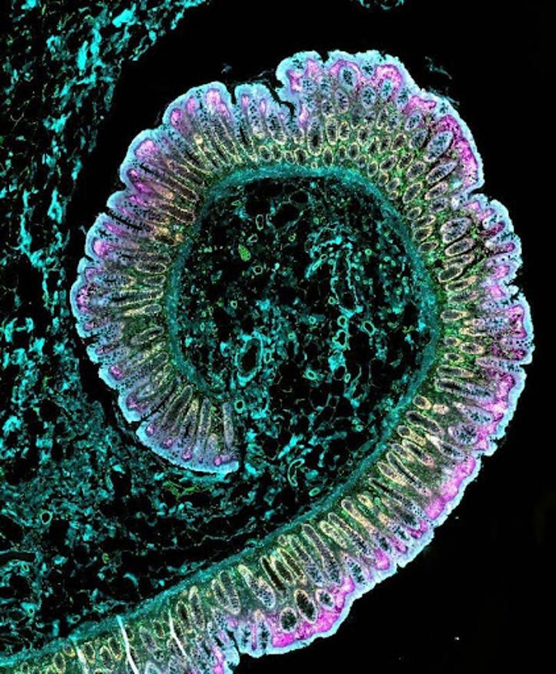
As the biotech and biopharma industries strive to develop more precise, personalized and effective therapies for complex diseases, researchers require access to deeper insights from cells, tissues and proteins. Spatial biology, the study of biomolecules and cells within their native tissue microenvironment, along with other advanced imaging techniques, is enabling scientists to better visualize, map and understand cell interactions in their natural locations than ever before.
Spatial biology and other cellular imaging processes are providing groundbreaking insights into research in critical disease areas, such as oncology, by enabling scientists to gain a more comprehensive understanding of biological processes. This view can include the interaction between immune cells, tissue-specific cells and blood vessels, as well as cancer cells when present. For example, researchers can clearly image immune cells and lysosomes as they invade and kill cancer cells (images 4, 11 and 12), enabling them to observe the biological processes underlying certain cancer immunotherapies and ultimately informing the discovery and development of new therapies. Cellular imaging, in combination with fields such as proteomics and genomics, can also help healthcare providers gain insights to predict and evaluate a patient’s response to a specific treatment in clinical settings.
Older cell imaging techniques involved analyzing isolated cells, which provided insight into cellular function but lacked a holistic view of cell organization and interaction. Additionally, older tissue analysis techniques could only reveal two to four distinct cell types in a single sample at a time, which also didn’t provide researchers with a comprehensive picture. Recent technological innovations have helped transform these analyses. Advancements, including clearer resolution, more complex data analysis methods and enhanced precision, have revolutionized our understanding of cell interactions in the human body, enabling the development of more targeted therapies.
Trisha Dowling
Other innovations in cellular imaging, like Thermo Fisher’s new Invitrogen EVOS S1000 Spatial Imaging System, help streamline imaging and analysis. Researchers can capture high-quality images of multiple targets simultaneously, reducing the need for multiple rounds of imaging and preserving sample integrity. Image 5 is a composite of more than 400 individual frames captured using the EVOS S1000 imaging system, providing a clear view of particular cell types and their structural arrangement within the tissue of a rat kidney. The EVOS S1000 can also provide researchers with images for multiple samples in only a few hours, saving researchers time.
Some solutions, like the EVOS S1000 imaging system, also help lower barriers to entry for labs looking to improve their cell imaging and analysis by simplifying data interpretation and offering compatibility with a range of reagents and antibodies, so labs may not need to buy a whole new suite of materials just for one new instrument.
Advanced cellular imaging and spatial biology techniques are shaping the future of research and development in new targeted therapies by providing researchers with a more precise and more in-depth understanding of how cells and tissues function and how they may respond to treatment.
Trisha Dowling is vice president of flow and imaging technologies, Thermo Fisher Scientific
Read the full article from the Source


